Figure 5.

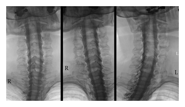
Cervical myelography (prone position). With the patient's head reclined, there is sufficient time to acquire images that show the cervical nerve roots in high detail without losing contrast. (Standard projections as Figure 3).

Cervical myelography (prone position). With the patient's head reclined, there is sufficient time to acquire images that show the cervical nerve roots in high detail without losing contrast. (Standard projections as Figure 3).