
Figure 8.
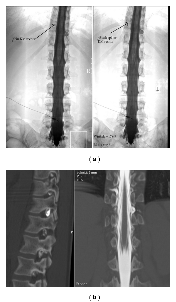
Spinal CSF leak causing subdural hematoma. (a) Left: contrast leakage to the left at the level of the D11 root. Right: 45 seconds later, contrast has flown around the dural sac and is exiting the spinal canal to the right. The dynamic series easily allows to study these flow dynamics and avoids misinterpretations. (b) Sagittal (left) and coronal (right) reformatted images from the subsequent myelo-CT show leakage in the left D11/12 foramen and contrast leakage to the right one segment above. This static study does not allow to exactly determine how contrast flows in and around the dural sac.