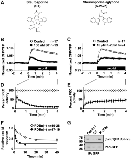
Figure 4. State-dependent inhibition by staurosporine and K-252c.
(A) The molecular structures for staurosporine (left) and staurosporine aglycone, K-252c (right). (B) Cellular PKC responses with (closed circles) or without (open circles) 100 nM staurosporine (ST). Application of 3 µM oxo-M (black box) is indicated. (C) Cellular PKC responses with (closed circles) or without (open circles) 10 µM K-252c. (D, E) Scaled PKC activities from (B) and (C). (F) Inhibition kinetics measured as oxo-M responses of PKC incubated with 100 nM staurosporine for the indicated times with (closed circles) or without (open circles) 100 nM PDBu. (G) Coimmunoprecipitation of Psd-GFP and (Δ2-31)PKCβII in the control or in the presence of 10 µM staurosporine or 10 µM K-252c.

