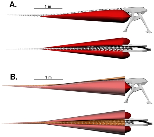
Figure 3. Lateral and dorsal views of the robustly modeled tail of Carnotaurus sastrei (MACN-CH 894).
(A) Digital reconstruction of the caudal and pelvic skeleton with M. caudofemoralis longus (red). (B) Complete digital reconstruction, with epaxial musculature (orange) and M. ilio-ischiocaudalis (pink) added.

