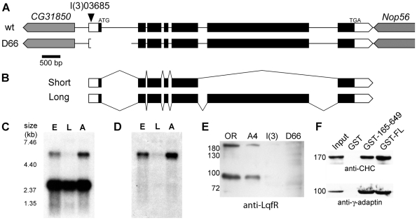
Figure 1. Characterization of Drosophila liquid facets-Related.
(A) Genomic region of lqfR, based on FlyBase release FB2008_07. lqfR is on the right arm of chromosome 3 and is oriented 5′ to 3′ towards the telomere. It occupies 7.7 kb and is flanked at the 5′ end by CG31850 and on the 3′ end by Nop56, both which are on the opposite (-) strand. The ATG-Start and TGA-Stop codons for the small splice product are shown. Within lqfR, white boxes represent UTR and black boxes represent ORF. Gene orientation is indicated by the pointed bars. The location of the P element insertion in lqfR 03685 is shown by the arrowhead. The lqfR D66 deletion is 1.1 kb. Scale bar, 500 bp. (B) The structure of the two predicted isoforms of lqfR is shown. Northern blots of embryonic, larval and adult mRNA probed with probes to lqfR exon 3–5 (C) and exon 6 (D). Marker sizes (in kb) are shown. (E) lqfR D66 mutants have undetectable amounts of LqfR protein. Western blot of lysates from 1/10 homozygous wild-type Oregon R (OR), lqfR +A4 (A4), lqfR 03685 (l(3)), or hypomorph lqfR D66 (D66) early pupae detected with crude rat anti-LqfR 3148-2 antiserum. (F) GST-pull down assays. GST alone, or GST fusions of full-length LqfR (GST-FL) or amino acids 165–649 (GST-165–649) were incubated with a soluble rat brain extract. Specifically bound proteins were detected with antibodies against clathrin heavy chain (CHC) or γ-adaptin. An aliquot of 1/10 of the brain lysate input was processed in parallel. Mr of protein standards is shown in kDA (E, F).

