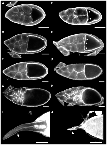
Figure 3. Developing egg chambers in lqfR mutants exhibit several morphological defects.
Stage-matched TRITC-phalloidin stained egg chambers from wild type (A,C,E,G,I) and lqfRD66 (B,D,F,H,J) ovaries. (A,B) Mid-stage 9; (C,D), stage 10A; (E,F) stage 10B; (G, H) stage 11 (H is slightly earlier in stage 11 than G, but it exhibits the cuboidal follicular epithelium and actin cytoskeleton of stage 11); (I,J) stage 14, anterior end only. Arrowheads show increased anterior actin in stage 9 and 10A (B,D) lqfR mutant egg chambers. Dorsal appendages (arrows). Anterior is at left in all panels. A–H are single confocal slices. I and J are z-stack confocal projections. Bars, 50 µm.

