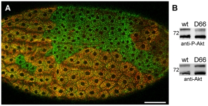
Figure 5. Liquid facets-Related mutants display a cell growth phenotype.
(A) Follicle cells of an early stage 14 egg chamber stained with anti-p120 (green) and anti-LqfR (red) showing two lqfRD66 clones. Image is a confocal z-stack projection. Anterior is to the left, dorsal is up. Bar, 50 µm. (B) Western blot of lysates from wild type and lqfRD66 adult females. The blot was first probed with anti-phospho-Akt (top), then stripped and reprobed with anti-Akt (bottom). Similar results were obtained when blots were probed first with anit-Akt and then anti-phospho-Akt1 (data not shown). Mr of the 72 kDa protein marker is shown.

