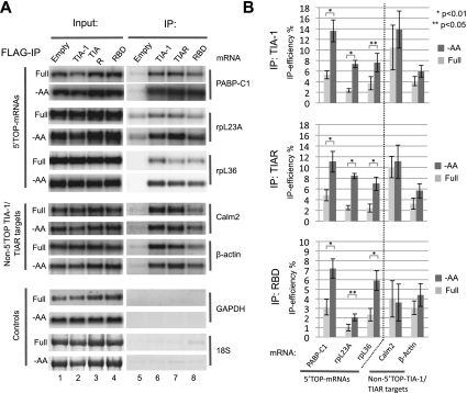
Figure 1.
Amino acid starvation stimulates assembly of TIA-1 and TIAR with 5′TOP mRNAs. (A) Northern blots comparing levels of mRNAs indicated on the right, in input fractions (lanes 1–4) versus pellet fractions (lanes 5–8) of immunopurified Flag-tagged TIA-1, TIAR, or C-terminally truncated TIA-1 (TIA-1 RBD) stably expressed in HEK293 cell lines, incubated for 2 h in either medium lacking amino acids (−AA) or with a full nutrient supplement (Full). (Lane 5) Immunopurification from cells expressing no Flag-tagged protein. Probed mRNAs are the 5′TOP-containing PABPC1, rpL23a, and rpL36 mRNAs (top three panels); calmodulin 2 and β-actin mRNAs, which lack 5′TOPs but contain TIA-1/TIAR-binding sites in their 3′ UTRs (middle two panels); and negative control GAPDH mRNA and 18S rRNA (bottom two panels). All mRNAs were detected with radiolabeled riboprobes, whereas 18S rRNA was detected using methylene blue staining. Input lanes represent 10% of the total RNA in immunoprecipitated samples. (B) Quantification of the data in A. Immunoprecipitation efficiencies were quantified as the percent of individual mRNAs found in pelleted material as compared with input fractions. Values and standard deviation were calculated from three independent experiments (n = 3). Dark-gray bars represent amino acid-starved cells (−AA), and light-gray bars represent cells in full medium (Full). P-values were calculated using Student's t-test (two tailed) and are indicated above the bars: (*) P < 0.01; (**) P < 0.05.

