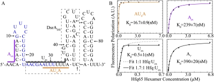
Figure 1.
Different Hfq-binding properties to selected segments on DsrA. (A) Schematic of the secondary structure of DsrA. Colored lines ([purple] Aex; [black] Uex; [orange] AU6A) along the structure show the fragments selected for investigation. The names of these fragments are colored accordingly. The nucleotides for rpoS base-pairing are colored in blue. DsrA domain II (DsrADII) consists of nucleotides in the dashed box. (B) FP of selected RNA fragments upon Hfq titration. A7 is a single-stranded poly(A) RNA segment. Equilibrium dissociation constants (Kd) of AU6A, Aex, and A7 were obtained by fitting to a 1:1 binding model. (Bottom left panel) Fitting of Uex in a 1:1 model (dashed lines) failed and ∼1.7:1 Hfq:Uex stoichiometry was required for good fitting of data points. Fitted curves are colored as in A for fragments from DsrA. Equilibrium dissociation constants are indicated in the graphs.

