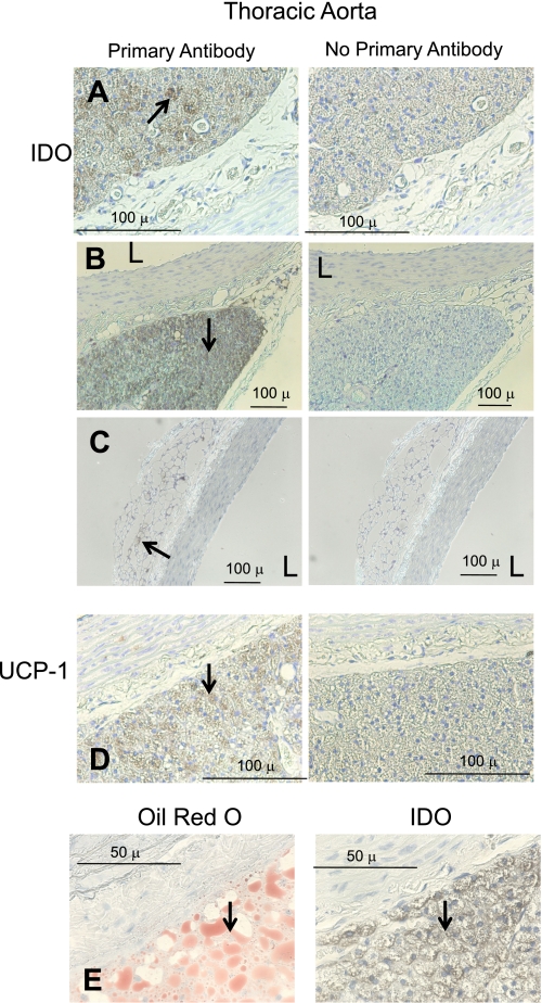
Fig. 2.
Immunohistochemical detection of IDO (A–C) and uncoupling protein-1 (UCP-1; D) in the thoracic aorta of a normal Sprague-Dawley rat. Left: with antibody; right: without antibody. A and B: representative staining of IDO in dense multilocular fat. C: IDO staining of the aorta with unilocular fat. D: UCP-1 staining. E: oil red O compared with IDO staining. Arrows point to regions of interest. Images are representative of four different animals. L, lumen of the vessel. Bars = 100 μm in A–D and 50 μm in E.

