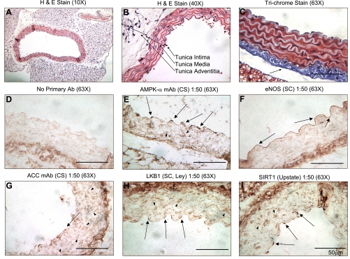
Fig. 6.
Hematoxylin and eosin (H&E), Masson's trichrome, and immunohistochemistry (IHC) of aortas from sedentary mice. Both A and B are H&E-stained aortas at different magnifications. The 3 layers of the aorta are defined in B. All Western blots were performed on protein lysates from the tunica intima [endothelial cell (EC) layer] and media [smooth muscle cell (SMC) layer] only. The aorta in C was stained with Masson's trichrome, which makes the nuclei appear black, cytosolic proteins appear red, and collagen appear blue. Note the thick layer of collagen attached to the tunica media (arrow). This collagen layer is immunoreactive with all antibodies, as is evident in D–I. D is a no primary antibody IHC control (i.e., only secondary antibody was used). AMPK-α (E), eNOS (F), ACC (G), LKB1 (H), and SIRT1 (I) were detected by IHC, respectively. Arrows point at positively stained EC, and arrowheads point at positively stained SMC. Above the IHCs, the primary antibody, company, dilution, and magnification of the micrograph are listed. CS, Cell Signaling; SC, Santa Cruz Biotechnology.

