Fig. 1.
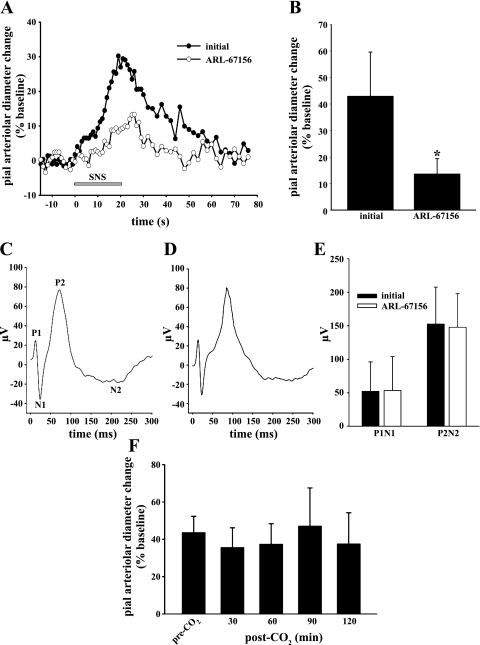
Sciatic nerve stimulation (SNS)-related data. A: representative time course of changes in pial arteriolar diameters associated with a 20-s SNS (gray bar). Each curve represents the average response measured in 2 animals, first in the absence (initial; ●) and, subsequently, in the presence of the ecto-pyrophosphatase/diphosphohydrolase blocker ARL-67156 (100 μM; ○). B: effect of ARL-67156 (100 μM) on SNS-evoked pial arteriolar dilations. Values are means ± SD. *P < 0.05 vs. initial; n = 5. Representative SNS-generated somatosensory-evoked potentials (SEPs) recorded from the contralateral cortical surface before (C) and after (D) ARL-67156 application are also shown. E: note that no differences in the SEP peak-to-peak amplitudes were observed, irrespective of whether the P1N1 or the P2N2 amplitude was measured. F: SNS-evoked pial arteriolar diameter percent increases from baseline measured before a CO2 challenge (Pco2, ∼70 mmHg for 3 min) and at 30-min intervals over the 2 h following CO2 exposure. This time control group displayed a remarkably consistent pial arteriolar response to SNS when comparing SNS-evoked pial arteriolar reactivities measured pre- and posthypercapnia. Results in E and F are means ± SD; n = 5.

