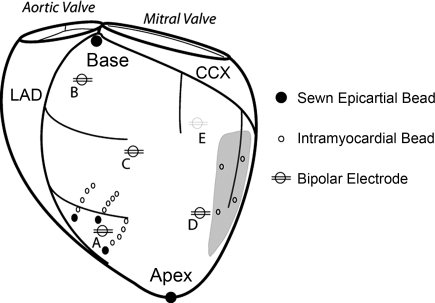
Fig. 1.
Experimental setup in nonfailing and failing hearts. Bipolar pacing electrodes were positioned on the left ventricle [i.e., in endocardial (ENDO) and epicardial (EPI) lead pairs] at the following locations: anterior apex (A), anterior base (B), anterolateral equator (C), lateral equator (D), and posterior coronary sinus (E; grey line). Note, in the nonfailing group the anterolateral equator location was replaced by a posterolateral equator location. Three columns of 4–6 intramyocardial markers (1–1.2 mm) were plunged into the anterolateral left ventricle and surface markers (1.7 mm) were sewn to the epicardial surface above each column. In the failing hearts, left ventricular scars (grey region) were present on the posterolateral wall and their locations were tracked with 3–4 gold markers (1.2–1.4 mm) implanted in the midwall. Left circumflex coronary (CCX) and left anterior descending (LAD) arteries are shown (black).

