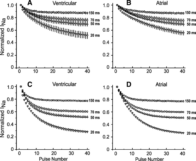
Fig. 2.
Abbreviation of the diastolic interval accentuates the development of use-dependent block of INa activated in atrial and ventricular cardiac myocytes by test pulses of constant duration in the presence of 10 μM (A and B) and 25 μM (C and D) ranolazine. A and C: inward INa recorded from ventricular myocytes during a train of pulses of 20-ms duration and diastolic intervals of 150, 70, 50, or 20 ms as a fraction of INa of the first pulse of the train (n = 6). B and D: INa recorded from atrial myocytes during a train of pulses of 20-ms duration and diastolic intervals of 150, 70, 50, or 20 ms as a fraction of INa of the first pulse of the train (n = 6). Data are means ± SD.

