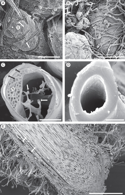
Fig. 3.
(A, B) Root hairs enmeshing sand grains (S) at the periphery of developing sandsheaths, approx. 10 and 75 mm, respectively, from the tip of a growing root like that in Fig. 1B. (C, D) Fractured root hairs, at approx. 5 and 100 mm, respectively, from a growing tip. The younger hair was living, with prominent nucleus (N), peripheral and transvacuolar cytoplasm (white arrows), and a relatively thin wall (black arrows). The older hair had developed a very thick wall before death. (E) Longitudinal face of dormant root showing aerenchyma formation in mid-cortex (arrows), and root hairs developed almost to the root tip. CSEM images. Scale bars: (A) = 165 µm; (B) = 375 µm; (C) = 5 µm; (D) = 5·5 µm; (E) = 1·2 mm.

