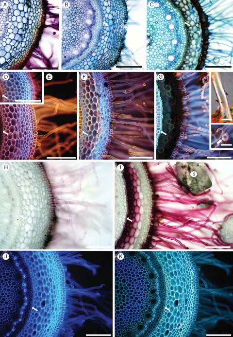
Fig. 6.
Fresh, hand-cut transverse sections of roots after removal of sand grains. The endodermis (arrow) is toward the left of each micrograph. Approximate distance (mm) of each section from the root tip is indicated. (A–C) Toluidine blue stain, at 10, 20, and 100 mm, respectively. (D–G) Rhodamine B-induced fluorescence at 10, 20, 100 and 180 mm, respectively. (Insets, G) Root hairs at 180 mm. A thin Sudan-positive layer (arrowheads) overlies thick, birefringent walls (upper inset G). Magnification of transverse view (arrow) of root hair (lower inset G). (H, I) Phloroglucinol/HCl (Wiesner test) at 20 and 100 mm, respectively. S, sand grain. (J, K) UV-induced autofluorescence of the same section at 20 mm, (J) in water and (K) in 0·1 m ammonium hydroxide. Scale bars: (A) = 200 µm; (B) = 300 µm; (C) = 250 µm; (D) = 275 µm; (E) = 175 µm; (F) = 200 µm; (G) = 150 µm; insets = 20 µm; (H) = 150 µm; (I) = 225 µm; (J, K) = 150 µm.

