Fig. 9.
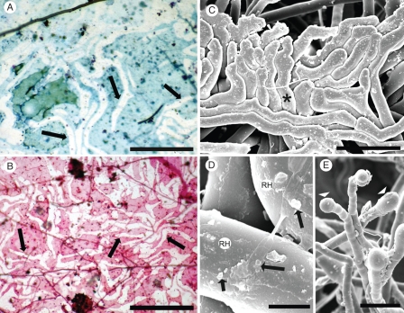
(A, B) Upper surface of mesh (0·03-μm pores) after 3 months in the field, along which a root grew, but no root hairs penetrated, and no sandsheath formed on the underside. After the root was removed gently, unstained root hair ‘ghosts’ (arrows) were revealed by ‘negative staining’ with Toluidine blue (A) and Schiff's reagent (B). Mesh between the ‘ghosts’ stained lightly by both methods. Fungal hyphae (arrowheads). (C) Root hairs, constrained by a hard surface (e.g. asterisk, Fig. 2B) form tissue-like closely adhering pads resembling the ‘ghost hair’ images in A and B. (D) Sparse deposits of possible mucilage and microbial colonies (arrows) on old hair surfaces (RH). (E) Possible exudates from young hairs. (C–E) CSEM. Scale bars: (A) = 140 µm; (B) = 140 µm; (C) = 75 µm; (D) = 6 µm; (E) = 80 µm.

