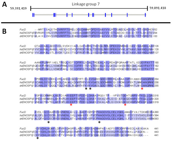
Figure 2.

The zebrafish homologue of hsENOSF1β. A: Genomic context of enosf1b. Blue bars are exons, grey dashed lines are introns. B: Alignment of enosf1b, hsENOSF1β, and FucD. Residues marked with a black asterisk are involved in FucD proton abstraction. Conserved residues marked with a red asterisk stabilize the magnesium ion required for FucD's catalytic mechanism.
