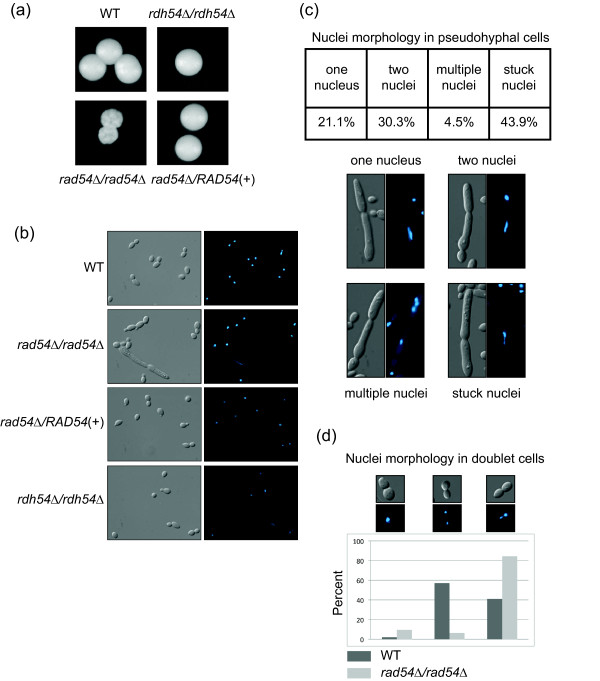
Figure 2.
Colony and cell morphology of rad54Δ/rad54Δ and rdh54Δ/rdh54Δ strains. A. Colony morphology after three days of growth on YPD is shown. B. DIC images and DAPI images of strains of the indicated genotypes. Note the aberrant cell and elongated nucleus in the rad54Δ/rad54Δ panel. C. Quantitation and examples of the nuclei morphology types seen in the ard54Δ/rad54Δ pseudohyphal cells. D. Quantitation and examples of the nuclei morphology in doublet cells in the WT and rad54Δ/rad54Δ cells.

