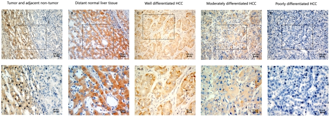
Figure 2. Immunohistochemical analysis of the LZAP protein expression in the primary hepatocellular carcinoma surgical specimens.
(A) and (F) Immunostaining of an HCC tumor and the adjacent non-tumorous area. (B) and (G) Normal liver tissue distant from the tumor, scored as LZAP (+++). (C) and (H) Well-differentiated HCC, scored as LZAP (++). (D) and (I) Moderately differentiated HCC, scored as LZAP (+). (E) and (J) poorly differentiated HCC, scored as LZAP (−). N: non-tumor tissue; T: tumor tissue (A–E with 200× magnification; F–J with 400× magnification).

