Figure 3.
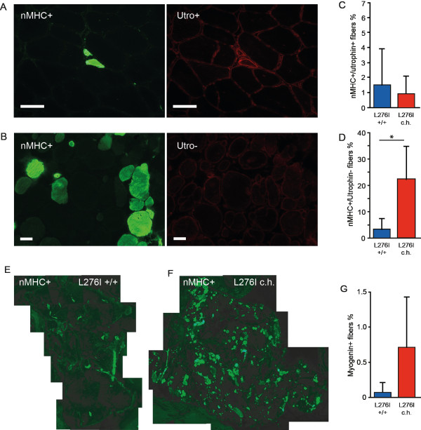
Muscle regeneration in patients with LGMD2I. (A) An illustration of newly regenerating fibers positive for neonatal myosin heavy chain (nMHC, green) and utrophin (red) (upper row), (B) Repair of mature fiber positive for nMHC and negative for utrophin (lower row). (C) The percentage of fibers positive for nMHC and utrophin did not differ between patients who were homozygous (n = 12) and those who were compound heterozygous (n = 5) for L276I. (D) The number of fibers positive for nMHC and negative for utrophin, as seen in fibers undergoing repair, was higher in compound heterozygous than in homozygous patients (P < 0.03). (E, F) nMHC expression in merged pictures of sections from (E) representative homozygous and (F) compound heterozygous patients. (G) The number of myogenin-positive nuclei trended towards an increase in compound heterozygous versus homozygous patients. Bar = 50 μm.

