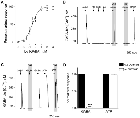
Figure 1. CHO cells stably expressing GABAB receptors are sensitive and reliable GABA biosensors.
Biosensors were loaded with Fura 2 and Ca2+ mobilization was measured in response to bath-applied GABA. A, Concentration-response relationships for GABA (symbols show mean ± s.e.m., N = 14 cells). B, Representative traces from a GABA biosensor showing that the biosensors do not respond to KCl depolarization (↓, KCl) or taste stimulation with a sweet/bitter mixture (↓, taste), and only minimally to acetic acid (↓, HAc). GABA-evoked Ca2+ responses (↓, GABA) were unaffected by removing extracellular Ca2+ (“0 Ca”, throughout the shaded area). Lastly, CGP55845 (“CGP”, 10 µM, present throughout the shaded area) reversibly blocked GABA-elicited biosensor responses. GABA, 100 nM; KCl, 50 mM. C, Representative traces from a GABA biosensor showing that biosensors respond to either GABA (100 nM) or ATP (10 µM), but CGP55845 (10 µM, present throughout the shaded area) only blocks responses elicited by GABA. D, Summary of data as shown in C. N = 6; ***, p<0.001, Student t-test.

