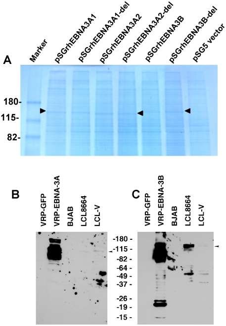
Figure 3. Expression of EBNA-3A and EBNA-3B in transfected and latently infected cells.
(A) Cos cells transfected with pSGrhEBNA3A1 and pSGrhEBNA3A1-del express EBNA-3A beginning at the first methionine without (lane 2) or with (lane 3) a deletion in the putative RBP-Jκ binding site, respectively. Cos cells transfected with pSGrhEBNA3A2 and pSGrhEBNA3A2-del express EBNA-3A beginning at the second methionine without (lane 4) or with (lane 5) a deletion in the putative RBP-Jκ binding site, respectively. Cos cells were transfected with pSGrhEBNA3B and pSGrhEBNA3B-del, with a deletion in the putative RBP-Jκ binding site (lanes 6 and 7, respectively). Cos cells were transfected with empty vector (pSG5, lane 8). (B) Detection of EBNA-3A in Vero cells infected with VRP-EBNA-3A, or in LCL8664 cells and in a rhesus monkey LCL (LCL-V). Arrow indicates location of EBNA-3A. (C) Detection of EBNA-3B in Vero cells infected with VRP-EBNA-3B, or in LCL8664 cells and LCL-V. Arrow indicates location of EBNA-3B. Additional bands noted in cells infected with VRP expressing EBNA-3A or -3B are likely due to overexpression of the protein.

