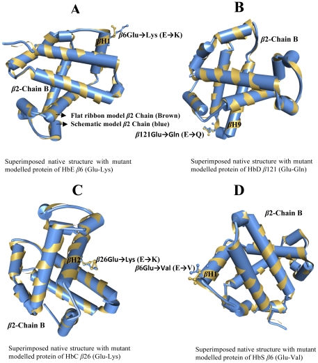
Figure 4. The superimpose alignments of crystal structure of human deoxyhaemoglobin native protein structure (4HBB) with mutant modelled protein i.e. HbE, HbD, HbC and HbS.
The structural modeling of proteins are flat ribbon model1 β2 Chain B (Brown) and Schematic model2 β2 Chain (blue). Four different amino acid mutations occur in the β2 Chain B, a) HbE contains β6Glu→Lys (E→K) located in the β Helix1, b) HbD contains β121Glu→Gln (E→Q) on located in the β Helix9, c) HbC contains β26Glu→Lys (E→K) in the β Helix2 and d) HbS contains β6Glu→Val (E→V) in the β Helix1.

