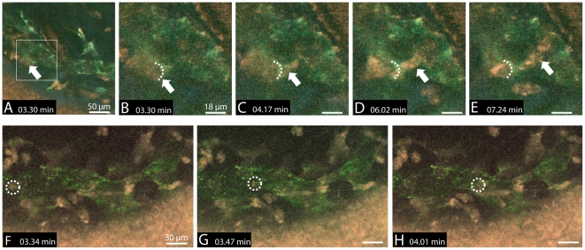
Figure 4. Intravital visualization of immune cell migration into and within a lymphatic vessel in a pathologically vascularized cornea.
A–E) Lymphatic vessel (green), crossing blood vessel (red), corneal epithelium (red) and stromal collagen fibrils (blue) are imaged simultaneously. An individual cell (arrow) migrates into the lymphatic vessel via a presumed gate (dotted line) that demonstrates an enhanced LYVE-1-antibody signal (compare to video S3). F–H) Rapid intravascular transport of a single cell (dotted circle).

