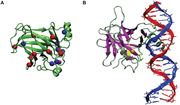
Figure 1. p53 DNA-binding core domain.
A) p53 DNA-binding domain mutations studied in this work. The zinc ion, destabilizing cancer mutations and stabilizing rescue mutations focused are depicted in purple, blue and red spheres, respectively. B) Different types of mutations in the p53 DNA-binding core domain. β-sheet residues, zinc-binding residues and DNA contact residues are depicted in purple, yellow and green, respectively. The zinc ion is depicted as an orange sphere.

