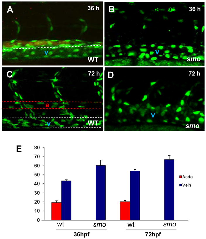
Fig. 3. The increase of venous cells is equavalent to loss of arterial cells in smo mutants.
(A–D) Confocal optics displaying individual nuclei of endothelial cells in the dorsal aorta (A,B stained with efnb2 antibody, the posterior cardinal vein and intersomitic vessels in wild-type embryos [Tg(fli:EGFP-nuc)] at 36 hpf (A) and 72 hpf (C),as compared to the increased endothelial nuclei in enlarged cardinal veins in smo mutants [smohi1640/smohi1640;Tg(fli:EGFP-nuc)] at 36 hpf (B) and 72 hpf (D). (E) Bar chart depicting number of arterial and venous cells in wild-type embryos compared to smo mutants. At 36 hpf, arterial cell number: 19.2± 1.7 (WT), 0 (smo); Venous cell number: 43.2±1.3 (WT), 60.3±2.4 (smo). At 72 hpf, arterial cell number: 20.2±1.3 (WT), 0 (smo); Venous cell number: 54±1.6 (WT), 66.8±3.4 (smo). Endothelial cells were counted from 12 lateral sections derived from 6 wild-type embryos and 6 smo mutants. Each section covers four segment lengths of intersomitic vessels along the dorsal aorta and the postertior cardinal vein in the middle trunk above the yolk extension region. a: aorta. v: vein.

