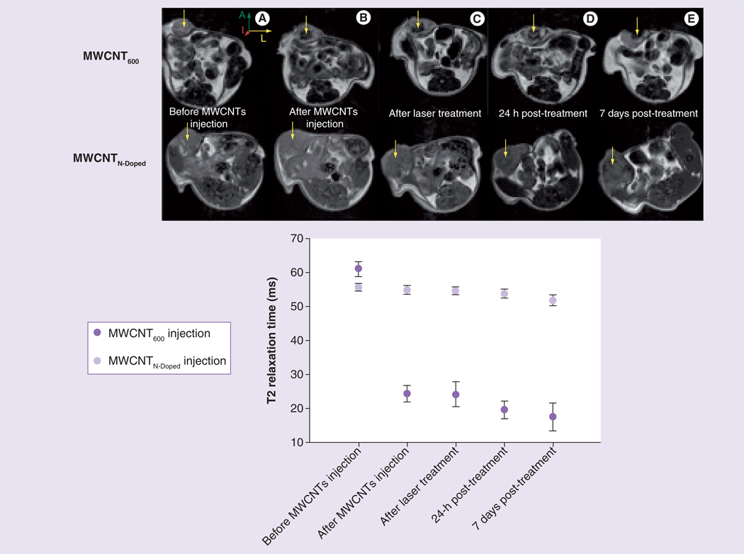
Figure 6. Magnetic resonance contrast properties of multiwalled carbon nanotubes are stable in vivo.
(A) Representative axial MR images of two breast tumors injected with either 100 µg MWCNT600 or MWCNTN-doped and subsequently exposed for 30 s to NIR laser. Arrows show the injection site. Images were collected at five timepoints: before injection, immediately after injection, immediately after laser treatment, 24 h after laser treatment, and 7 days after laser treatment (laser power: 3 W/cm2, 1064 nm wavelength). (B) T2 relaxation measurement of the tumor region was acquired using multislice multipulse sequence at the five different time points described in (A).
A: Anterior; I: Inferior; L: Left; MR: Magnetic resonance; MWCNT: Multiwalled carbon nanotube; N-Doped: Nitrogen-doped.

