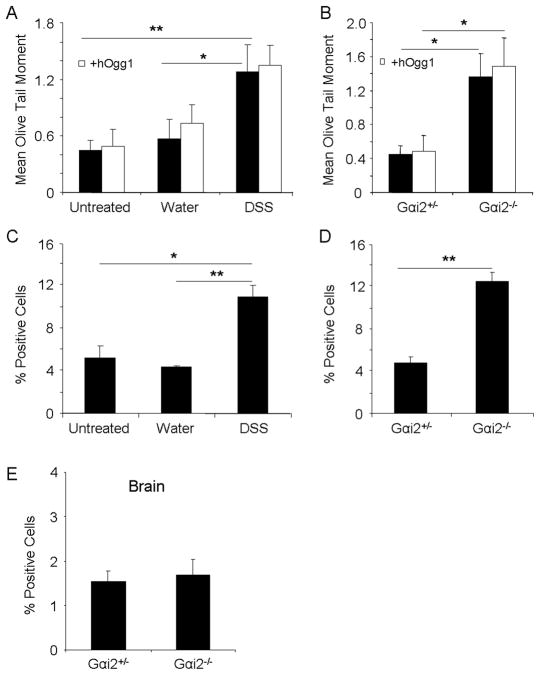
Fig. 4.
DNA damage to hepatocytes and brain in colitic mice. A, C. DNA damage as measured by alkaline comet assay with and without hOgg1 incubation, and percent positive cells for γ-H2AX foci formation, respectively, in wildtype mice treated with DSS or water for two cycles. B., D. DNA damage as measured by alkaline comet assay with and without hOgg1 incubation, and percent positive cells for γ-H2AX foci formation, respectively, in Gαi2−/− or age-matched Gαi2−+ − mice. E. Percent positive cells for γ-H2AX foci formation in transverse sections of the brain in Gαi2+/− and Gαi2−/− mice. *: p<0.05, **: p<0.01 by one way ANOVA with Dunn’s multiple comparison test. Error bars represent SEM of 5–7 mice per group.

