Figure 5.

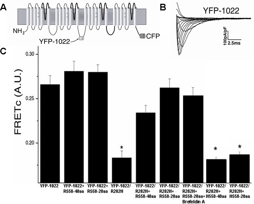
FRET in Normal and Mutant Channels A. Diagram of FRET construct containing CFP fused to the C-terminus of hNav1.5 and YFP inserted into the Domain II-III linker at amino acid position 1022. B. Currents elicited from the FRET construct are similar to WT currents. C. FRETc values for YFP-1022 (n=43) were similar to YFP-1022+R558-40aa (n=35), YFP-1022+R558-20aa (n=39), YFP-1022/R282H+R558-40aa (n=43), YFP-1022/R282H+R558-20aa (n=46), and YFP-1022/R282H+R558-20aa+Brefeldin A (n=34). FRETc of YFP-1022/R282H (n= 45) alone, YFP-1022/R282H+H558-40aa (n=36), and YFP-1022/R282H+H558-20aa (n=42) were significantly smaller than WT. *P<0.05 compared to WT.
