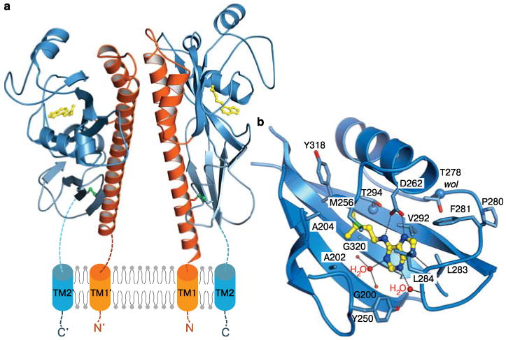
Figure 1.

AHK4 binds cytokinins with its membrane-distal PAS domain. a, Ribbon diagram of the sensor domain homodimer (residues 126-391). The N-terminal stalk helix and the dimerisation interface is shown in orange, the membrane-distal cytokinin binding domain in dark-blue, and the membrane-proximal PAS domain is in light-blue, respectively. Disulphide bridges are depicted in green. One molecule of iP (in yellow) binds per AHK4 monomer. b, Close-up of the cytokinin binding pocket complexed with iP (in bonds representation).
