
Figure 1.
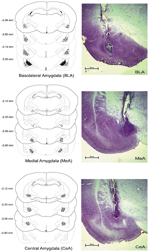
Schematic of coronal sections of the rat brain showing the placements of the tips of the cannulae for all rats that received infusions of HU-210, AM251 or URB597 into the basolateral nucleus of the amygdala, the central nucleus of the amygdala and the medial amygdala. Representative histological pictures of infusions into the amygdala nuclei are adjacent to placement diagrams.