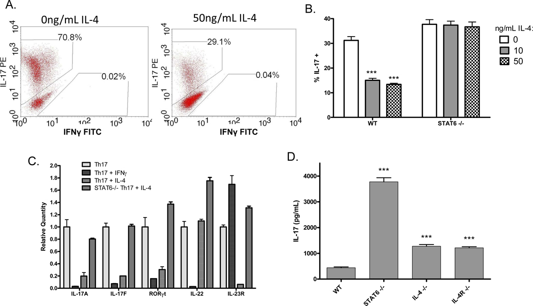
Figure 3. IL-4 suppresses a different subset of Th17 genes than IFNγ, and the effect of IL-4 is mediated by STAT6.
Naïve CD4+ T cells from wildtype or STAT6-deficient mice were cultured under Th17-skewing conditions for five days, followed by two days rest and two days re-stimulation in the presence or absence of 50ng/mL IL-4 or IFNγ. (A) Cells were treated for six hours with PMA, ionomycin and brefeldin A, and IL-17 expression was measured by ICS. (B) Cells were treated for six hours with PMA, ionomycin and brefeldin A, and IL-17 expression was measured by ICS. Error bars represent SEM of triplicate cultures. *** p<0.001 vs. 0ng/mL IL-4 by one-way ANOVA. (C) RNA was collected and IL-17A, IL-17F, RORγt, IL-22 and IL-23R expression was measured by real-time PCR with primers and probes from Applied Biosystems. Data is normalized first to â-actin, the internal control, and then to the matched sample without IL-4 or IFNγ. Error bars represent SEM of triplicate PCRs. (D) Augmented IL-17 expression in STAT6-deficient mice. Spleen cells from wildtype BALB/c, STAT6 knockout, IL-4 knockout, or IL-4R knockout mice were stimulated in vitro with anti-CD3. Five days later supernatant was collected and IL-17 expression was measured by ELISA. Bars represent mean SEM of triplicate samples. ***p<0.001 vs. wildtype by one-way ANOVA.

