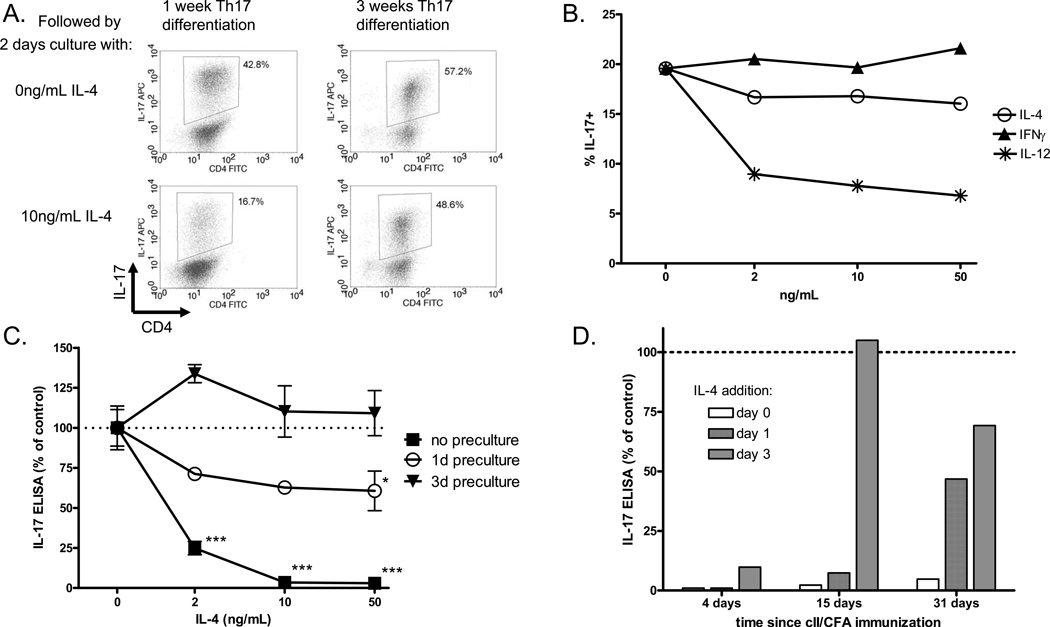
Figure 8. Th17 cells become resistant to suppression by IL-4 both in vitro and ex vivo.
(A.) Th17 cells were generated by five days stimulation and two days rest as described, followed by two days re-stimulation in the presence or absence of IL-4 and ICS for IL-17. Alternatively, cells were cultured for two more rounds of five days Th17 stimulation and two days rest, for a total of three weeks of culture. After three weeks, the cells were re-stimulated for two days in the presence or absence of IL-4, followed ICS for IL-17. (B) Th17 cells cultured for three weeks to induce maturation were re-stimulated for two days with increasing concentrations of IL-4, IFNã or IL-12, followed by ICS for IL-17. (C) DBA mice were immunized i.d. with cII/CFA and spleens were collected two weeks later. Total spleen cells were re-stimulated with heat-denatured collagen for zero, one or three days, and then collected, washed and re-plated with collagen and increasing concentrations of IL-4 for two days. Supernatants were collected and IL-17 was measured by ELISA. Bars represent mean +/− SEM of triplicate culture samples, normalized to 0ng/mL IL-4. *p<0.05, *** p<0.001 vs. 0ng/mL IL-4 by one-way ANOVA. (D) DBA mice were immunized with cII/CFA and spleens were collected 4, 15 or 31 days laters. Spleen cells were re-stimulated with collagen and 50ng/mL IL-4 was added on day 0, 1 or 3. Supernatants were collect on day five and IL-17 was measured by ELISA. IL-17 expression is normalized to the sample with no IL-4.

