
Figure 2.
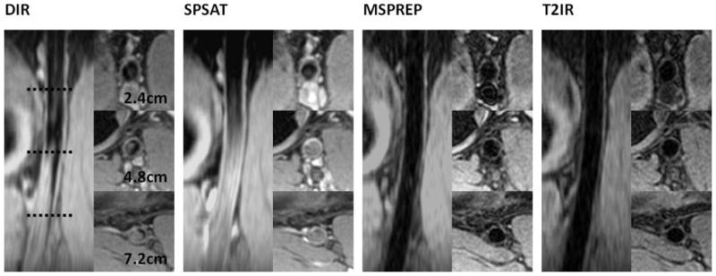
Axial source and coronal reformatted vessel wall images of the popliteal artery obtained with the four BB techniques in the same subject. This case followed the general trends observed in this study.
Official websites use .gov
A
.gov website belongs to an official
government organization in the United States.
Secure .gov websites use HTTPS
A lock (
) or https:// means you've safely
connected to the .gov website. Share sensitive
information only on official, secure websites.
Axial source and coronal reformatted vessel wall images of the popliteal artery obtained with the four BB techniques in the same subject. This case followed the general trends observed in this study.