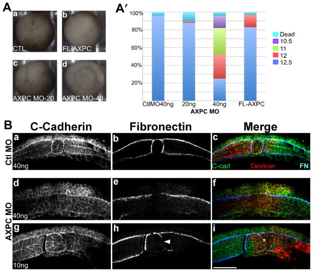
Figure 3.
Loss of AXPC expression perturbs notochordal morphogenesis
A. 2-cell stage embryos were injected bilaterally in the dorsal marginal zone with 40ng control morpholino (a), 20 or 40ng AXPC morpholino (c,d) or 1ng AXPC mRNA (d) and cultured until uninjected controls reached stage 12.5. Stage was determined by blastopore size and percentage was calculated by: # in given stage/total # embryos (A′, n>50). B. Whole mount immunofluorescence of bisected embryos injected with either control morpholino (CtlMO, a–c) or AXPC morpholino-2 (AXPC MO2, d–i). Sections were stained to detect C-cadherin to outline cell borders (a,d,g) and fibronectin to delineate tissue boundaries (b,e,h). Dextran tracer can be observed as red in c,f, and i. Arrowhead in h marks a localized loss of FN staining. Confocal images acquired at 200X magnification. Scale bar = 150μm.

