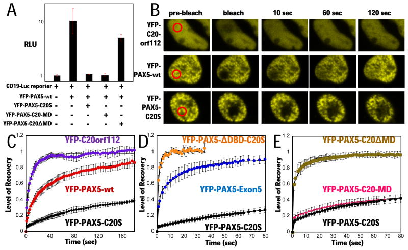
Figure 2. PAX5-C20S suppresses wt PAX5 activity and causes extremely slow nuclear mobility in living cells.
(A) A YFP-PAX5-wt vector and the luciferase reporter were either transfected or co-transfected with vectors for YFP-PAX5-C20S, YFP-PAX5-C20-MD, or YFP-PAX5-C20ΔMD and luciferase assayed (RLU). (B) Fluorescence micrographs from FRAP assays in 293T cells. Red circled areas were photobleached regions, and fluorescence signal in each area was sequentially measured. FRAP assays of (C) YFP-PAX5-C20S (black circles, N=4, t1/2 ~ 200 sec), YFP-C20orf112 (purple circles, N=4, t1/2 ~ 3 s) and YFP-PAX5-wt (red circles, N=7, t1/2 ~ 24 s) in 293T cells, (D) YFP-PAX5-ΔDBD-C20S (orange circles, N=3, t1/2 ~ 1 s), YFP-PAX5-C20S (black circles, N=4, t1/2 ~ 200 s), and YFP-PAX5-Exon5 (blue circles, N=3, t1/2 ~ 4 s) in 293T cells, (E) YFP-PAX5-C20ΔMD (gold squares, N=6, t1/2 ~ 1 s), YFP-PAX5-C20-MD (pink diamonds, N= 7, t1/2 ~200 s), and YFP-PAX5-C20S (black circles, N=5, t1/2 ~200 s) in H1299 cells. Error bars here and in subsequent FRAP assays represent standard error of the mean.

