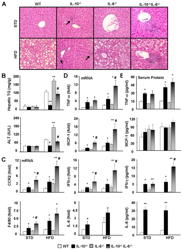
Figure 2. HFD-induced steatosis, liver inflammatory response and injury are exacerbated in IL-10−/−IL-6−/− dKO mice compared with IL-10−/− mice.
WT, IL-10−/−, IL-6−/−, IL-10−/−IL-6−/− mice were placed on a HFD or STD for 12 weeks. (A) H&E staining of liver sections. (B) Liver triglyceride and serum ALT levels. (C, D) Real-time PCR analyses of hepatic inflammatory markers CCR2 (for monocytes) and F4/80 (for macrophages) (C) and hepatic cytokines (D). (E) Serum levels of cytokines. *P<0.05, **P<0.01 in comparison with corresponding WT group; # P<0.05 in comparison with corresponding IL-10−/− group.

