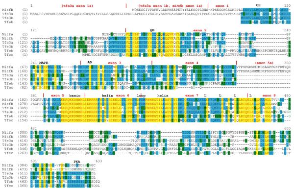
Figure 1. Multiple sequence alignment of the zebrafish MiT family.
Alignment of amino acids sequences derived from ClustalW is shown. Red text on yellow background indicates highest similarity, followed by blue on cyan, black on green, and green on white. Vertical red lines indicate exon boundaries, and the basic region helix-loop-helix domain, and leucines comprising the leucine zipper are indicated. Abbreviations: CH, charged helical domain; QB, glutamine-rich, basic domain; MAPK, consensus MAP kinase phosphorylation site; AD, transcriptional activation domain; PKA, consensus cAMP-dependent protein kinase phosphorylation site.

