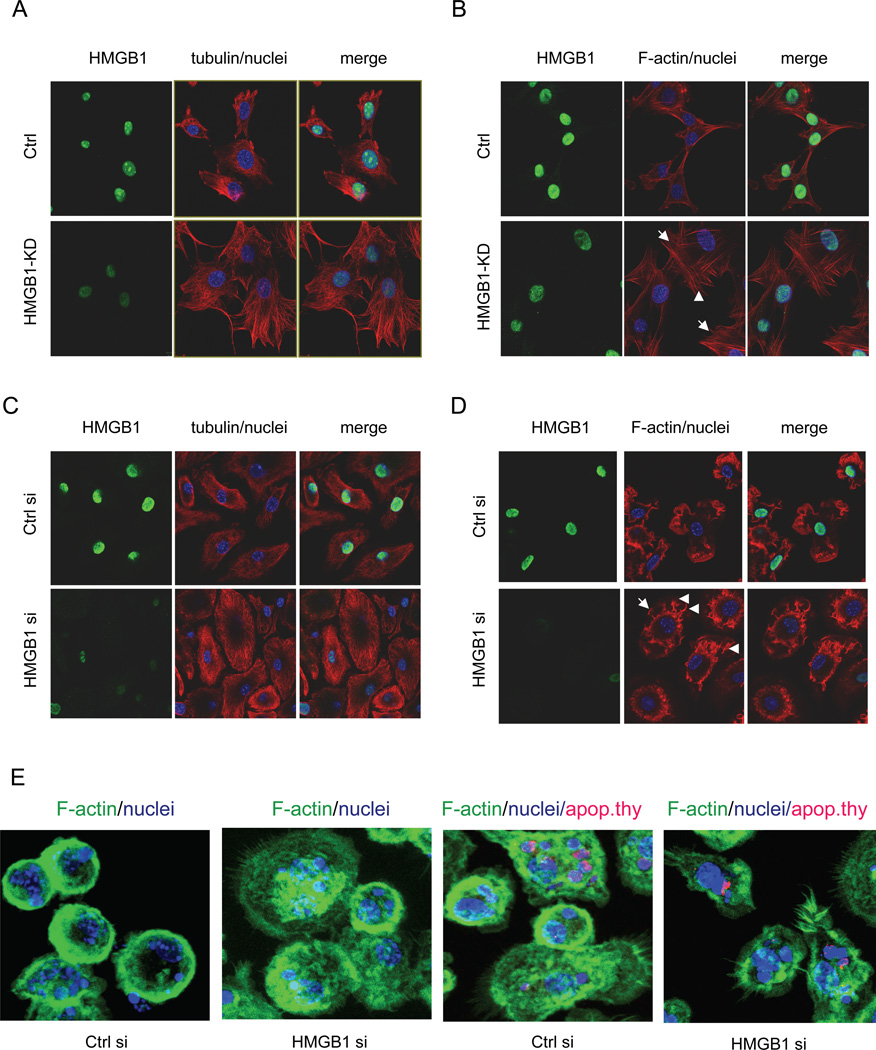
Figure 5. Enhanced cytoskeletal rearrangement is present after HMGB1 knockdown.
(A) NIH3T3 cells transfected with pBabe-H1-HMGB1, but with minimal HMGB1 knockdown (Ctrl), and NIH3T3 cells with HMGB1 knockdown (HMGB1-KD) were fixed and permeabilized. The cells were then incubated with primary anti-α-tubulin and anti-HMGB1 antibodies, followed by incubation with fluorescence conjugated secondary antibodies. DAPI was used to stain nuclei. (B) Experiments were performed as in (A), except that the primary anti-α-tubulin antibody was replaced with phalloidin for staining of F-actin. The arrows show microspikes and filapodia. (C–D) Murine BMDMs were transfected with control siRNA or siRNA to HMGB1. At 72 hours after transfection, the cells were fixed, permeabilized and stained as A and B for imaging. (E) Murine BMDMs were transfected with control siRNA or HMGB1 siRNA. At 72 hours after transfection, the cells were incubated without or with Phrodo-labeled apoptotic thymocytes (red fluorescence) for 1h. The cells were then washed thoroughly to remove uningested thymocytes and fixed, permeabilized and stained with FITC-conjugated phalloidin for identification of F-actin (green fluorescence).

