
Fig. 6.
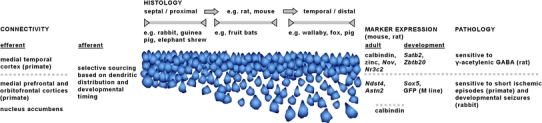
Examples of particularly interesting histoarchitectural, connectional, neurochemical and pathological findings that distinguish superficial and deep pyramidal cells of CA1. The cell layer has been drawn to resemble the different appearances of the layer along both the septotemporal and proximodistal axis in mouse and rat. In other species much of CA1 is dominated by a histoarchitectural phenotype that encompasses only a narrow segment of the variations seen in rat and mouse