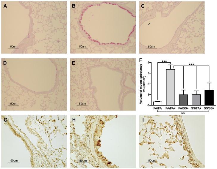
FIGURE 7. SS exposure suppresses the allergen-induced airway mucus, and SPDEF formation.
Lung sections (5 μm) were stained with AB-PAS for mucus, and SPDEF as described in the Methods. Representative histologic photomicrographs (40x) of the lung sections show the mucus-producing cells in pink, and SPDEF in brown. Mucus staining: (A), FA/FA; (B), FA/FA+; (C), FA/SS+; (D), SS/FA+; (E), SS/SS+; (F), Volume density(Vs) of AB-PAS-stained mucosubstances in the mucosal surfacein the lungepithelium (n = 5/group; bars represent the mean ± SD; *** p ≤ 0.001; and NS = not significant). SPDEF staining: (G), FA/FA; (H), FA/FA+; (I), SS/FA+ (n =3/group); + indicates Af-sensitized.

