
FIGURE 2.
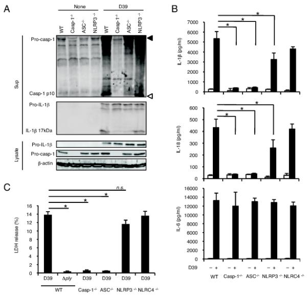
ASC is essentially required for caspase-1 activation in S. pneumoniae infected macropahges. Adherent PECs from C57BL/6 WT, caspase-1−/−, ASC−/−, NLRP3−/− or NLRC4−/− mice were left uninfected or infected with S. pneumoniae D39 or the Δply strain at a MOI of 10 for 24 h as described in Fig. 1, and culture supernatants and cell lysates were then collected. A: Culture supernatants and cell lysates were subjected to Western blot analysis as described in Fig. 1. Filled and open triangles indicate MW markers of 47 kDa and 9 kDa, respectively. B: Levels of IL-1β, IL-18 and IL-6 in the culture supernatants were determined by ELISA. C: LDH release from D39- or Δply-infected macrophages derived from each mouse strain was determined by the LDH activity in the culture supernatants, and the data are expressed as percent LDH release. All of the experiments were repeated more than three times. The results are presented as the mean and SD of triplicate assays. Tests for statistical significance were performed by using one-way ANOVA followed by the Bonferroni’s test. n.s., not significant. *, p < 0.05.