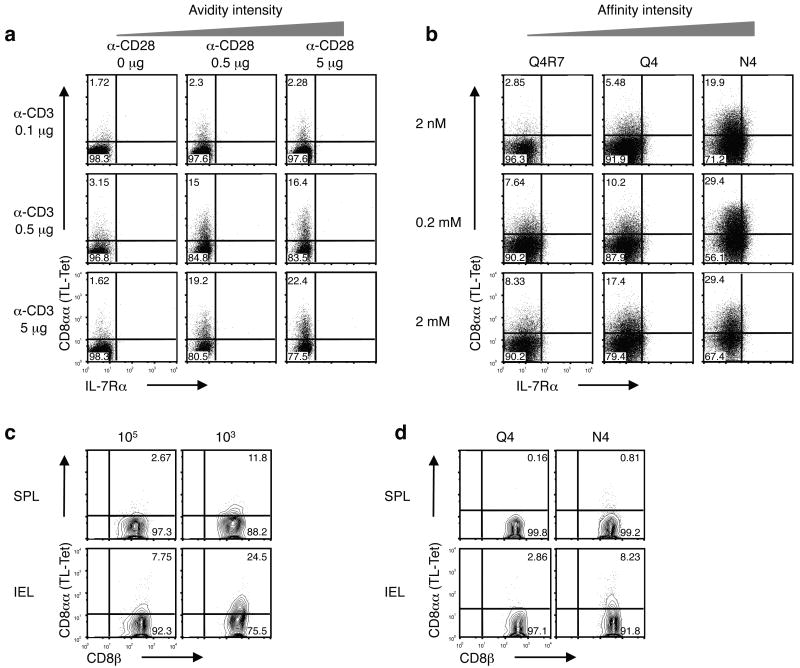
Figure 4. CD8αα expression correlates with the intensity of TCR activation.
(a) Total splenocytes were cultured in the presence of graded concentration of soluble anti-CD3 and anti-CD28. CD8αα expression, as measured by TL tetramer staining, was analyzed 3 d after in vitro culture. Representative data on gated CD8+ T cells are shown. IL-7Rα expression is also depicted. Three independent experiments were performed. (b) Naïve OT-I cells were cultured with artificial APC (MEC.B7) in the presence of graded concentration of OVA257-264 SIINFEKL (N4) or altered peptide ligands (Q4R7 and Q4). CD8αα expression was detected 2 d after in vitro culture. Three independent experiments were performed. (c) 1 x10⁵ or 1 × 10³ sorted naïve Ly5.1+ CD8+ OT-I cells were transferred into B6 recipient mice. 1 d after transfer, mice were orally infected with 1 × 10⁹ ActA- Lm-OVA. 7 d p.i., CD8αα expression was analyzed on Ly5.1+ CD8+ OT-I cells from the spleen and IEL (representative data from a single mouse is shown, n = 4 mice per group). (d) 5 × 10⁴ naïve Ly5.1+ CD8+ OT-I cells were transferred into WT recipient mice. 1 d after transfer, mice were orally infected with 2 × 10⁸ WT Lm-Q4OVA or Lm-N4OVA. 7 d p.i., CD8αα expression was analyzed on donor OT-I cells from the spleen and IEL (representative data from a single mouse is shown, n = 5 mice per group). Data are representative of three (a, b) and two (c, d) independent experiments.

