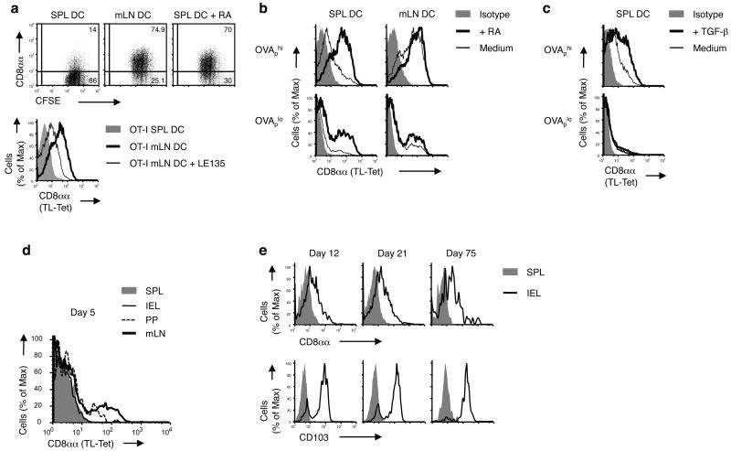
Figure 6. Retinoic acid promotes the affinity-based accumulation of CD8αα+ CD8αβ T cells in the intestine.
(a) OT-I cells were stimulated by OVAp-loaded DCs from SPL or mLN of WT mice with or without 100 nM RA (dot plots) or with LE135 (histogram) in vitro for 3 d and CD8αα expression was analyzed. Data are representative of five independent experiments. (b,c) OT-I cells were stimulated by SPL or mLN DCs pulsed with OVAp (high, 1 nM; low, 0.01 nM) in the presence or absence of 100 nM RA (b) or 5 ng/ml TGF-β (c) in vitro for 3 d and CD8αα expression was analyzed. Data are representative of more than five independent experiments. (d,e) 0.5 × 10⁶ CD8+ OT-I cells isolated from naïve Ly5.1+ OT-I+ Rag-/- mice were adoptively transferred into B6 recipient mice. 1 d after transfer, mice were orally infected by 0.5 × 10⁹ ActA- Lm-OVA. CD8αα expression was measured on gated donor OT-I cells from the SPL, mLN, PP and IEL, 5 d p.i. (d). CD8αα and CD103 expression is shown on gated donor OT-I cells from the spleen and IEL on days 12, 21 and 75 p.i. (e). Representative data from two to three mice per group are shown. At least three independent experiments were performed.

