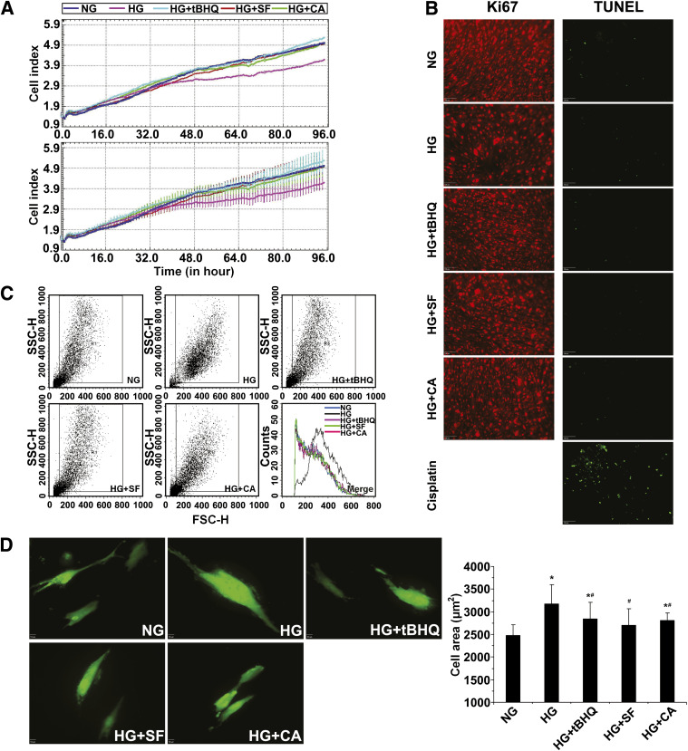
FIG. 6.
High glucose–mediated mesangial cell growth inhibition and hypertrophy can be reversed by activation of Nrf2. A: Cell growth of HRMCs incubated in NG, HG, HG+tBHQ, HG+SF, or HG+CA DMEM media was monitored in real-time for 96 h (upper panel = average; lower panel = average with error). B: Cell death and proliferation were assessed by transferase-mediated dUTP nick-end labeling (TUNEL) assay (positive control is treated with cisplatin at 18 μmol/L for 24 h) or Ki67 immunolabeling. HG media induced cell growth inhibition, which was alleviated by coculture with an Nrf2 activator. C: Cell size of HRMCs incubated in NG, HG, HG+tBHQ, HG+SF, or HG+CA DMEM media for 96 h was measured by forward light scatter/flow cytometry. Incubation in HG media induced cellular hypertrophy that was reduced with an Nrf2 activator. D: Cell area is reported from GFP-transfected HRMCs incubated in NG, HG, HG+tBHQ, HG+SF, or HG+CA DMEM media for 48 h. Total cell area increased with HG conditions but was significantly reduced in the presence of an Nrf2 activator. Data in D are expressed as mean ± SD (n = 100). *P < 0.05 compared with NG group. #P < 0.05 Nrf2 activators compared with HG alone. FSC-H, forward scatter. SSC-H, side scatter. (A high-quality color representation of this figure is available in the online issue.)

