
FIG. 3.
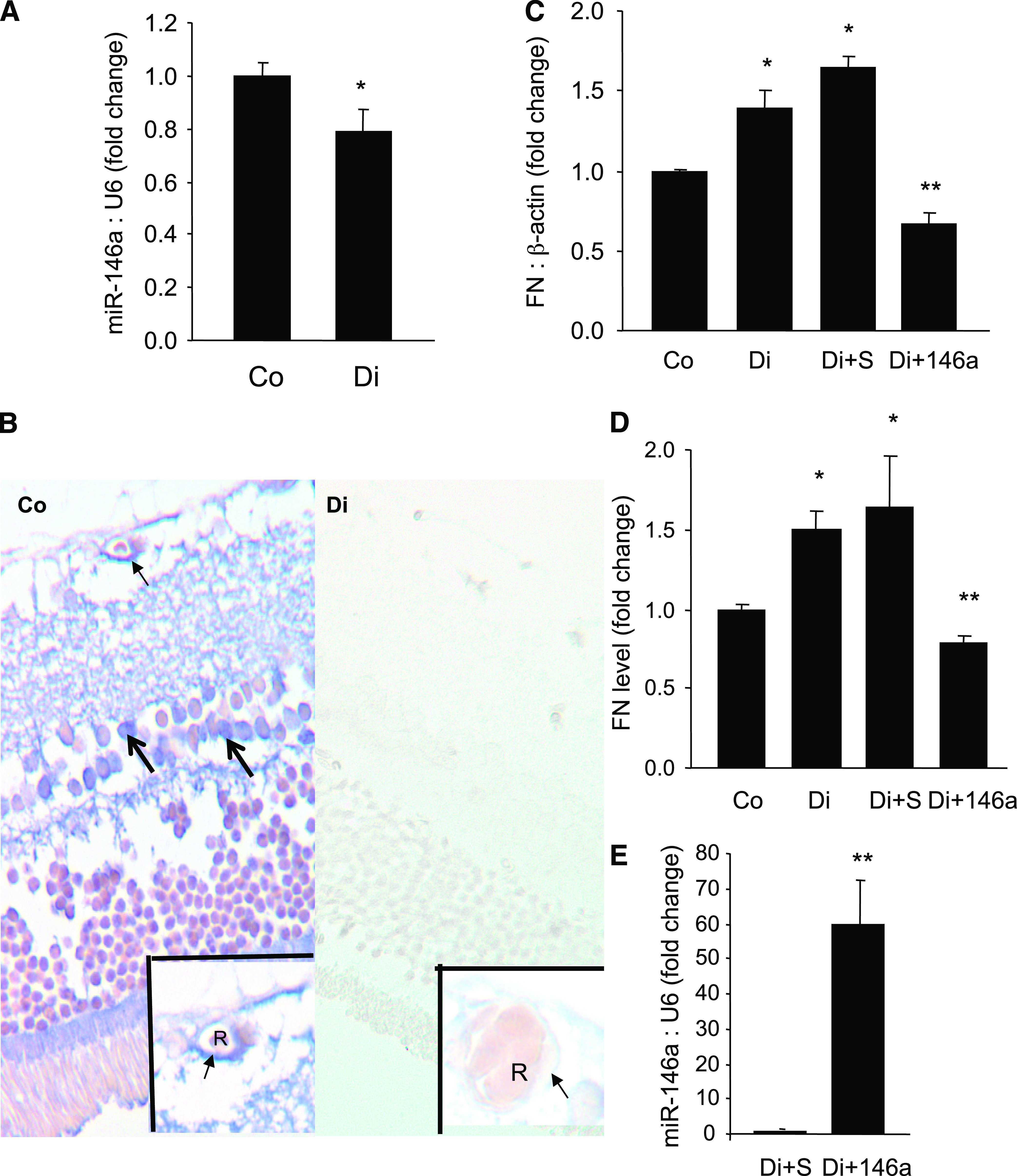
miR-146a–mediated alteration of retinal FN. A: Poorly controlled diabetes caused a reduction of rat retinal miR-146a levels. B and C: Representative LNA-ISH study of retinal tissues in a control (Co) and a diabetic (Di) rat retina showing localization of miR-146a (blue chromogen) in the retinal capillaries (arrow) and in the cells of inner nuclear layer (large arrow), possibly both in the glial and neuronal elements. Insets show an enlarged view of the capillary with miR-146a localization (arrow) in a control rat and reduced level of endothelial miR-146a in the retina of a diabetic rat (arrow). Diabetes-induced augmented FN mRNA (C) and protein levels (D) (as measured by ELISA) in the rat retina can be prevented by intravitreal miR-146a mimic (but not by scrambled [S] mimics) injection. E: Efficiency of intravitreal delivery as demonstrated by increased retinal miR-146a expression after an intravitreal injection of miR-146a mimic compared with scrambled mimic. *Significantly different from control; **significantly different from diabetic or diabetic plus scrambled. miRNA levels are expressed as a ratio of RNU6B (U6) normalized to control or diabetic plus scrambled (E). mRNA levels are expressed as a ratio to β-actin and normalized to control. Alkaline phosphatase was used as chromogen (blue) with no counterstain in LNA-ISH. R, red blood cell. (A high-quality digital representation of this figure is available in the online issue.)