FIG. 3.
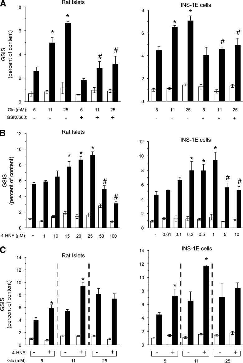
4-HNE mimics high glucose amplification of insulin secretion. A: Rat islets and INS-1E cells were incubated with RPMI-1640 medium containing the indicated glucose levels in the absence or presence of 1 μmol/L GSK0660 for 48 and 24 h, respectively. GSIS was evaluated by 1-h static incubations at 3.3 mmol/L glucose (white bars), followed by a 1-h incubation at 16.7 mmol/L glucose (black bars). Insulin secretion is presented as percent of insulin content. Glc, glucose. Results are mean ± SEM, n = 3. *P < 0.05 for the difference of stimulated secretion (16.7 mmol/L glucose) compared with untreated controls maintained at 5 mmol/L glucose. #P < 0.05 for differences from the respective GSK0660-free incubation. B: Isolated rat islets and INS-1E cells were incubated for 48 or 24 h, respectively, in standard RPMI-1640 medium (11 mmol/L glucose) in the absence or presence of increasing concentrations of 4-HNE. GSIS was measured as in A. The vehicle ethanol, at a 1:1,000 dilution, did not affect GSIS in either β-cell preparation. Insulin secretion is presented as percent of insulin content. Results are mean ± SEM, n = 3. *P < 0.05 relative to the stimulated secretion of vehicle-treated controls. #P < 0.05 relative to the maximal stimulatory level. C: Rat islets and INS-1E cells were incubated for 48 and 24 h, respectively, at 5, 11, or 25 mmol/L glucose in the absence or presence of 4-HNE (15 or 1 µmol/L, respectively) followed by GSIS analysis. Results are mean ± SEM, n = 3. *P < 0.05 for difference from the respective untreated controls, exposed to the same glucose concentration.

