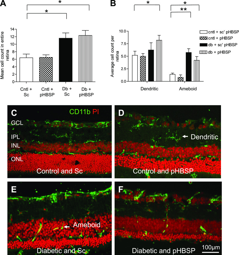
FIG. 4.
pHBSP and microglial activation in the diabetic retina. Retinal microglia were labeled using CD11b immunoreactivity in retinal sections and visualized using confocal microscopy. Cntl, control; Sc, scrambled pHBSP; and Db, diabetic. A: Mean cell counts of CD11b-positive cells were taken from three separate points within the central retina. There was a significant increase in microglial numbers after 7.5 months of diabetes (*P < 0.05; **P < 0.01). pHBSP (10 μg/kg) had no significant effect on this diabetes-related increase (P > 0.05). B: After subdividing the total number of microglial cell counts, there is a significant difference in the number of dendritic (nonactivated) and amoeboid (activated) cells between control and diabetic rats (*P < 0.05). Compared with diabetic rats treated with the scrambled peptide, there are more dendritic microglia and fewer amoeboid in the retinae of the diabetic rats that received the scrambled pHBSP (**P < 0.01 and *P < 0.05). Data are mean ± SEM; n = 6 per group. Sc’pHBSP, scrambled pHBSP. C–F: Images of CD11b-positive cells: control and Sc (C), control and pHBSP (D), diabetic and Sc (E), and diabetic and pHBSP (F). (A high-quality color representation of this figure is available in the online issue.)

