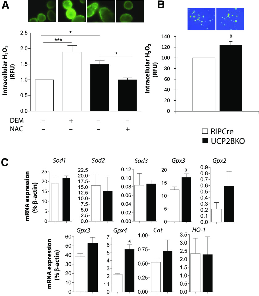
FIG. 3.
Higher intracellular ROS levels and antioxidant gene expression in UCP2BKO islets. A: Fluorescent microscopy was used to image intracellular H2O2 levels in islets isolated from RIPCre and UCP2BKO mice that were cultured overnight and incubated with the ROS-sensitive fluorescent dye CM-H2-DCFDA, in the presence of 11 mmol/L glucose. Islet intracellular H2O2 levels were manipulated by incubation of RIPCre islets with (+) or without (−) the pro-oxidant DEM (5 mmol/L) or UCP2BKO islets with (+) or without (−) the antioxidant NAC (0.2 mmol/L). Data shown are expressed as the fold-change over RIPCre islets. Representative fluorescent microscopy images of each condition are shown above each bar. n = 11–16 islets from 4 mice/genotype. *P < 0.05, ***P < 0.0001. B: Measurement of intracellular H2O2 in β-cells selected from dispersed islet cells. H2O2 was measured as in A. A total of 20–30 larger cells (i.e. β-cells) were selected for each coverslip. n = 5–7. C: Quantitative PCR was used to quantify the expression of several antioxidant genes as well as the oxidative stress-responsive HO-1 in isolated islets. Data shown are expressed as a percentage of β-actin mRNA expression levels for n = 3–7. *P < 0.05. (A high-quality digital representation of this figure is available in the online issue.)

